Extracting magnetic information from Kerr Microscopy images
ResearchComplete
Sole author. BSc Artificial Intelligence dissertation, University of Manchester. Awarded 76% (first class).
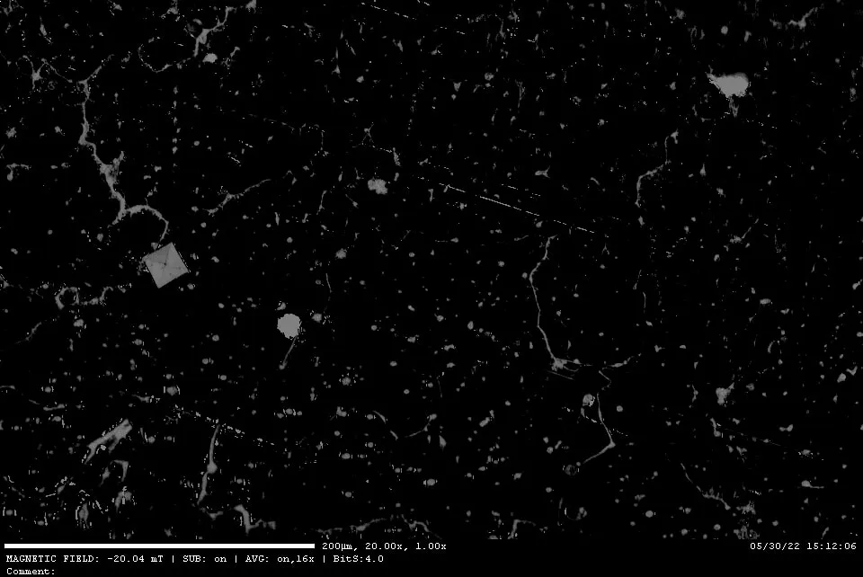
An automated image-processing pipeline for Kerr microscopy that improves image analysis and the extraction of magnetic data, with a web application interface to support research.
My final-year project at the University of Manchester, awarded 76% (first class). The project involved computer vision and image processing.
It develops an automated image-processing pipeline for Kerr microscopy to improve image analysis and the extraction of magnetic data, with a web application interface to support research.
Documents
Extracting magnetic information from Kerr Microscopy images
Dissertation58 pagesNot peer-reviewed
- Open in a new tab: Extracting magnetic information from Kerr Microscopy images (PDF)
- Download (PDF, 2.2 MB): Extracting magnetic information from Kerr Microscopy images
Cite Extracting magnetic information from Kerr Microscopy images
Plain text
Lennon, J. (2025). Extracting magnetic information from Kerr Microscopy images [BSc Artificial Intelligence dissertation, University of Manchester]. https://joshlennon.com/documents/kerr-microscopy-dissertation.pdf
BibTeX
@thesis{lennon2025extracting,
  author      = {Lennon, J},
  title       = {{Extracting magnetic information from Kerr Microscopy images}},
  type        = {BSc Artificial Intelligence dissertation},
  institution = {University of Manchester},
  year        = {2025},
  note        = {Not peer-reviewed},
  url         = {https://joshlennon.com/documents/kerr-microscopy-dissertation.pdf},
}
Media
Authorship
This is my own work.
Supervised by Prof Thomas Thomson.